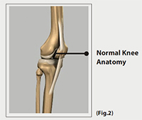
Articular cartilage is a complex avascular (no blood supply) tissue which consists of cells called chondrocytes suspended in a collagenous matrix. It appears as a smooth, shiny, white tissue at the ends of the bones which come in contact with each other to form a joint.
This cartilage is subjected to the normal wear and tear and may sometimes get damaged because of injury causing pain and impaired function.
Function
Articular cartilage reduces the friction when the bones glide over each other and makes the movements smooth and enables the joint withstand weight. Alternately, it acts as a shock-absorber.

Causes
Articular cartilage injuries occur as a result of sports injury (direct blow) or progressive degeneration (wear and tear). Degeneration of the cartilage occurs as a progressive loss of structure and function of the cartilage. The process begins with softening of the cartilage which then progresses to fragmentation. As the articular cartilage lining is lost, the underlying bone has no protection against the normal wear and tear and it starts breaking down leading to osteoarthritis. The risk factors that can contribute to osteoarthritis include twisting injuries, abnormal joint structure, instability of joints and inadequate muscle strength.
The symptoms of cartilage injuries include:
- Pain and swelling of the knee joint
- Locking of the knee
- Catching or giving way
Diagnosis
Your doctor will perform a physical examination to look for altered range of motion, swelling, and alignment of the bones. As cartilage is uncalcified it does not show up in X-rays. A high quality MRI often required and arthroscopy is used as the final determination to what technique may be best used.
Treatment
Initial treatment includes physical therapy, anti-inflammatory medications, and steroid injections. Surgery to restore articular cartilage may be considered for patients with large articular lesion or if conservative treatment fails. Articular cartilage repair is performed to provide relief from pain, improve range of motion, slow the progression of the damage, and delay the option of joint replacement surgery.
Indications for surgery
Surgery is usually not necessary when the cartilage defect is small and asymptomatic. Defects which are smaller than 2 cm can be treated arthroscopically and larger defects may require transplantation of cartilage from other areas of the joint. Most of the cartilage restoration procedures are done using an arthroscope.
The surgical procedures for cartilage restoration include:
- Microfracture: Microfracture involves creating numerous tiny holes in injured joint surface using a special tool, called ‘awl’. The holes are made in the bone under the cartilage, called as subchondral bone. This creates a new blood supply to the cartilage which stimulates the growth of new cartilage.
- Drilling: This procedure is similar to microfracture where multiple holes are created in the injured joint area using a surgical drill or wires.
- Abrasion arthroplasty: This procedure is similar to drilling but involves use of high speed burs to remove the damaged cartilage.
- Autologous chondrocyte implantation (ACI) – Ii is a two step procedure, where healthy cartilage cells are removed from the non-weight bearing joint, grown in the laboratory and then implanted in the cartilage defect during the second procedure. During this procedure a patch is harvested from the periosteum, a layer of thick tissue that covers the bone and is sewn over the defected area using fibrin glue. The new cartilage cells are then injected under the periosteum into the cartilage defect to allow the growth of new cartilage cells.
- Osteochondral autograft transplantation: In this procedure, plugs of cartilage is taken from the non-weight bearing areas of knee, from the same individual and transferred to the damaged areas of the joint. This method is used to treat smaller cartilage defects since the graft which is taken from the same individual will be limited.
- Osteochondral allograft transplantation: In this procedure healthy cartilage tissue or a graft is taken from a donor from the bone bank and transplanted to the area of cartilage defect.
Following cartilage replacement your doctor may recommend physical therapy to help restore mobility to the affected joint.
 Call for an Appointment


